Medical Imaging for Diagnosis, Treatment
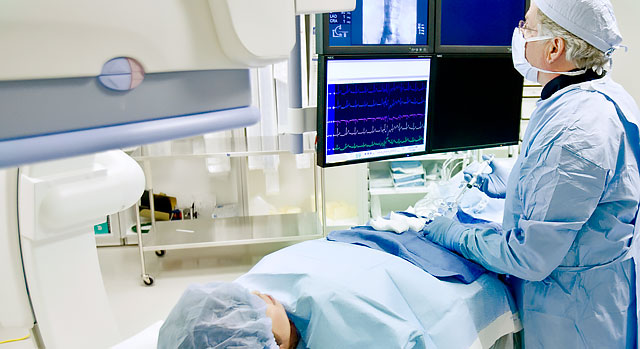
Radiology is the area of medicine that uses X-rays, radioactive tracers, magnetic waves and ultrasonic waves to obtain detailed images of the inside of the body. Doctors use these images to detect illnesses and injuries and to develop treatment plans.
Several imaging tests are available at Valley Hospital Medical Center.
X-ray
An X-ray image is produced when a small amount of radiation passes through the body to expose sensitive film on the other side. The ability of X-rays to penetrate tissues and bones depends on the tissue's composition and mass. The difference between these two elements creates the images. The chest X-ray is the most common radiologic examination. Contrast agents, such as barium, can be swallowed to highlight the esophagus, stomach and intestine, and are used to help visualize an organ or film.
CT
Computed tomography (CT) shows organs of interest at selected levels of the body. They are the visual equivalent of bloodless slices of anatomy, with each scan being a single slice. CT examinations produce detailed organ studies by stacking individual image slices. CT can image the internal portion of organs and separate overlapping structures precisely. The scans are produced by having the source of the X-ray beam encircle or rotate around the patient. X-rays passing through the body are detected by an array of sensors. Information from the sensors is processed by a computer and then displayed as an image on a video screen.
MRI
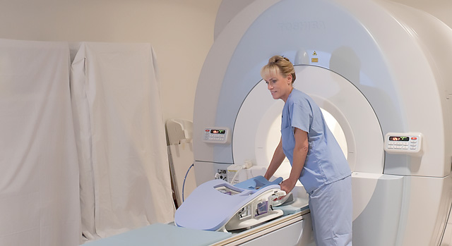
Like CT, magnetic resonance imaging (MRI) produces images that are the visual equivalent of a slice of anatomy. MRI, however, is also capable of producing those images in an infinite number of projections through the body. MRI uses a large magnet that surrounds the patient, radio frequencies and a computer to produce its images. As the patient enters an MRI scanner, his or her body is surrounded by a magnetic field up to 8,000 times stronger than that of the earth.
The scanner subjects nuclei of the body's atoms to a radio signal, temporarily knocking select ones out of alignment. When the signal stops, the nuclei return to the aligned position, releasing their own faint radio frequencies from which the scanner and computer produce detailed images of the human anatomy.
Patients who cannot undergo a MRI examination include those people dependent upon cardiac pacemakers and those with metallic foreign bodies in the brain or around the eye.
FibroScan®
FibroScan non-invasively measures the stiffness of your liver by capturing and calculating the speed of a shear wave as it travels through the liver. This detection of stiffness may be used as an aid to clinical management of liver disease.
When performed as part of an overall evaluation, FibroScan provides valuable information for healthcare providers that might otherwise only be available from a liver biopsy.
Nuclear medicine
Nuclear medicine is a medical specialty that uses safe, painless and cost-effective techniques to image the body and treat disease. Nuclear medicine imaging is unique in that it documents organ function and structure, in contrast to diagnostic radiology, which is based upon anatomy. It is a way to gather medical information that may otherwise be unavailable, require surgery or necessitate more expensive diagnostic tests.
Nuclear medicine is used in the diagnosis, management, treatment and prevention of serious disease. Nuclear medicine imaging procedures often identify abnormalities very early in the progression of a disease, long before some medical problems are apparent with other diagnostic tests. This early detection allows a disease to be treated sooner in its course, when there may be a more successful prognosis.
Nuclear medicine uses very small amounts of radioactive materials, or radiopharmaceuticals, to diagnose and treat disease. Radiopharmaceuticals are substances that are attracted to specific organs, bones or tissues. The radiopharmaceuticals used in nuclear medicine emit gamma rays that can be detected externally by either gamma or PET cameras. These cameras work iwith computers to form images that provide data and information about the area of body being scanned.
The amount of radiation from a nuclear medicine procedure is comparable to that received during a diagnostic X-ray. Today, nuclear medicine offers procedures that are helpful to a broad span of medical specialties, from pediatrics and cardiology to behavioral health.
There are nearly 100 different nuclear medicine imaging procedures available and all major organ systems which can be imaged by nuclear medicine.


